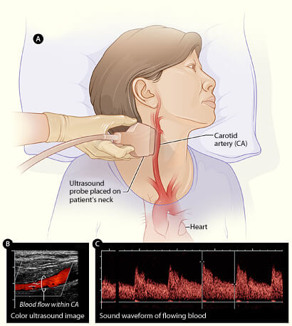
Obesity is a global, ever-increasing epidemic. In 2020, there are already 650 million people with this disease. Obesity leads to the development of cardiovascular risk factors – hypertension, dyslipidemia, diabetes mellitus, sleep apnea, and independently increases cardiovascular diseases and mortality. The distribution of adipose tissue, the so-called visceral and ectopic obesity, is an independent indicator of adverse events. The main pathophysiological changes are systemic inflammation, endothelial dysfunction, insulin resistance. Treatment is medical and surgical and builds on changes in lifestyle, nutrition and exercise regimen, with promising results in terms of coronary disease, heart failure and atrial fibrillation.
Keywords: obesity, visceral, ectopic, cardiovascular risk factors
Our perception of obesity has been shaped over the years by beloved cartoon,
television and literary characters, by classic images in the visual arts, and
many times it evokes affection and indulgence. Regardless of any aesthetic value we attach to it, obesity is a polyetiological disease, which is expressed by a body mass index (BMI) ≥30 kg/m2 and through various mechanisms leads to an increase in cardiovascular diseases and mortality. In the case of NSTEMI in a young person, obesity turns out to be the most important risk factor, along with smoking. The higher the BMI, the earlier the NSTEMI will occur. An increase in BMI by 1 unit increases the risk of ischemic stroke by 4%, and the risk of hemorrhagic stroke by 6%. A 10 kg weight gain increases the risk of coronary heart disease by 12%, systolic blood pressure by 3 mmHg, and diastolic blood pressure by 2.3 mmHg. (1) Therefore, the definition of obesity should not be limited to BMI alone, but mainly to cardiovascular and metabolic complications in the body. Abdominal and visceral obesity have a stronger relationship with cardiometabolic complications. (2) Therefore, in the risk assessment we include waist circumference and waist-hip ratio as easily measurable indicators. With the imaging methods CT and MRI, the distribution of fat can be differentiated, mainly subcutaneous or visceral, the latter being more closely correlated with risk.
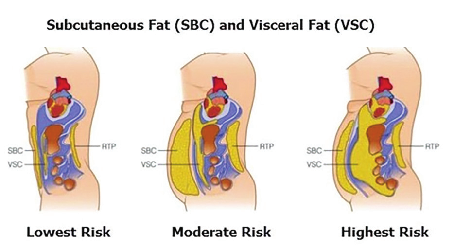
A category of “metabolically healthy” obese individuals has been identified, in whom, despite high BMI and waist circumference, visceral fat distribution is smaller. Visceral obesity is accompanied by hepatic steatosis – another form of fat distribution associated with the same risk factors, namely arterial hypertension, dyslipidemia and diabetes mellitus. Another ectopic fat accumulation is pericardial and epicardial fat. Pericardial fat is the total amount of fat in the pericardial sac, assessed by CT. Epicardial fat is visceral fat between the outer layer of the myocardium and the visceral leaf of the pericardium and shows a relationship with waist circumference, indicators of insulin resistance, dyslipidemia, blood pressure and especially with sleep apnea.
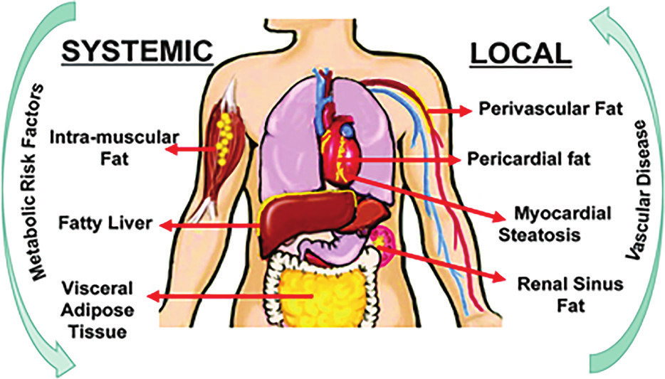
Dietary regimen and physical activity reduce visceral and pericardial adiposity and thus risk, regardless of whether they lead to weight change. (3)
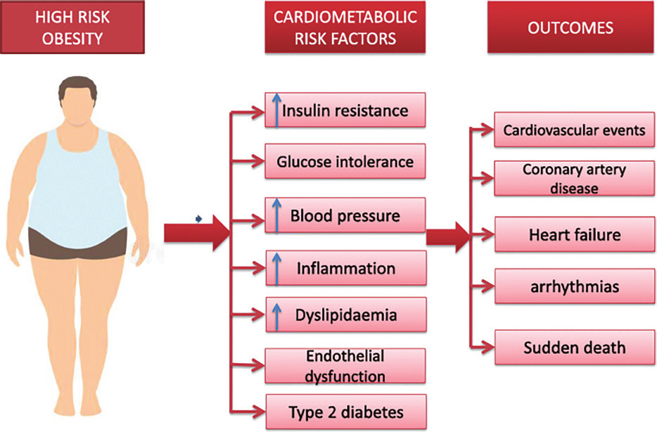
Main pathophysiological mechanisms of metabolic and cardiovascular diseases
Lipid accumulation and adipose tissue enlargement in the absence of changes in blood supply leads to hypoxia, NIF1a is activated, which in turn is the cause of fibrosis initiation and develops in visceral adipose tissue. Oxidative stress and loss, in turn, are the cause of reduced adiponectin production and increased secretion of leptin, resistin, proinflammatory adipokines and cytokines, TNFa, IL-6, IL-8, free fatty acids from adipocytes. Thus, local perivascular disease develops into systemic. If adiponectin has a protective effect on myocardial hypertrophy, cardiac fibrosis, atherosclerosis, then leptin is the cause of insulin resistance, endothelium dysfunction, activation of the RAAS and the sympathetic nervous system. All these processes are the basis of atherosclerosis and coronary disease, arterial hypertension, diabetes mellitus and heart failure. For the development of HF with reduced chest wall compliance and accumulated cardiovascular risk factors. (4) The role of lipotoxicity caused by high levels of free fatty acids must be added, which, in addition to myocytes, damage the pancreas (islet apoptosis), liver (steatosis and fibrosis), muscles (insulin resistance) and kidneys (fibrosis and renal failure). Adipose tissue also exerts mechanical compression, e.g. peripharyngeal, such as that in obstructive sleep apnea. Compression of the kidneys by accumulated fat in and around them is a possible mechanism for increasing blood pressure. High intrarenal pressure reduces tubular flow, with the most compressive segment being the loop of Henle in the medulla. This leads to compensatory increased reabsorption of sodium and chlorine in the ascending limb and, accordingly, less NaCl reaches the macula densa, glomerular filtration, renin and angiotensin II increase, which potentiates Na-reabsorption of the arterial and this pressure. In recent years, great importance has been given to the red microbiota. In obesity, lactobacillus, E. coli, fecal bacterium, which are gram-negative, rich in LPS, dominate. An obesogenic diet, poor in fiber and cellulose, leaves bacteria without a substrate for the final products of fermentation. Low levels of butyrate are the cause of intestinal permeability disruption, translocation of bacteria and lipopolysaccharide in the circulation, activation of Toll like R 4, systemic cycle, activation of macrophages and formation of atherosclerotic plaques. A diet rich in meat is a source of choline, L-carnitine and betaine, metabolized by bacteria to TMA (trimethylamine), which is converted to TMAO (trimethylamine N-oxide) in the liver. High levels of TMAO via NF-kB lead to the expression of genes with proinflammatory and, accordingly, oxidative stress effects. TMAO causes platelet hyperactivation and thrombosis. There is evidence that red dysbiosis induces and maintains high blood pressure levels through the systemic syndrome, vasoactive metabolites such as serotonin, dopamine, norepinephrine, indoxyl sulfate, etc. (5)
Pathophysiology of sudden death and atrial fibrillation
Obesity is the most common non-ischemic cause of SCD – every 5-unit increase in BMI carries a 16% higher risk. Potential mechanisms include LVH, QT-prolonged interval, ventricular extrasystoles and autonomic imbalance. A common finding is the fragmented QRS complex, a surrogate for heterogeneous conduction, which, together with fibrosis, is an independent predictor of SCD. The association with atrial fibrillation is even stronger. Obesity is responsible for 1/5 of cases of atrial fibrillation. Mechanisms include left atrial remodeling, fibrosis, expression of endothelin receptors, epicardial fat infiltrating the underlying myocardium with a change in conduction. (6)
Clinical guidelines in history and examination
Obesity is not a hidden disease when perceived only as BMI. Most patients feel healthy. The presence of appetite and weight gain is always perceived as a “good sign”. Let us not forget the “obesity paradox”. (9) Complications are difficult to diagnose if we do not actively look for them. CT should not be done to determine visceral/subcutaneous obesity with a larger waist circumference. It is sufficient to test for insulin resistance, diabetes mellitus, and abdominal ultrasound for hepatic steatosis. (10)
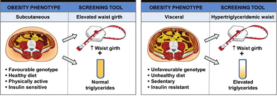
Measuring blood pressure with a standard cuff at the level of the arm in a patient with extreme obesity is a real challenge and leads to an overestimated result. Some researchers recommend doing this at the level of the forearm, and the values will be even higher. (11) When reading the ECG, it is taken into account that the heart is elevated from the diaphragm, located more horizontally in the mediastinum, distant from the precordial electrodes, so the Cornell score is used to assess LVH: R aVL + S V3 >35 mm in men and >25 mm in women. The heart rate is high, usually with Q waves in the lower leads, suspicious for a previous myocardial infarction – especially since angina complaints are common. All forms of obstructive and non-obstructive coronary disease are characteristic. Microvascular dysfunction may be suspected in younger patients with few or no traditional cardiovascular risk factors, more often in women, but even then it is associated with a higher risk. (12) Complaints of shortness of breath, fatigue, and edema are common, and may be symptoms of HF with preserved or reduced EF, noting that BNP levels are inversely related to BMI. Left ventricular concentric remodeling, right ventricular dilatation and dysfunction, and pericardial constriction with interventricular dependence are more common in HFpEF, especially with more epicardial fat. Coronary artery disease is a cause of systolic dysfunction and HFrEF. The history should include guiding questions about sleep apnea and rhythm disturbances, as well as screening for atrial fibrillation.
Obesity Treatment
The Look AHEAD study showed that participants who lost ≥10% of their weight had a significant reduction in cardiovascular risk. This was associated with changes in blood sugar, blood pressure, and lipids.
Individuals who achieved ≥10% weight loss were 6 times less likely to have an atrial fibrillation episode. Current treatment is not simply BMI-centered, but is aimed at reducing CV risk.
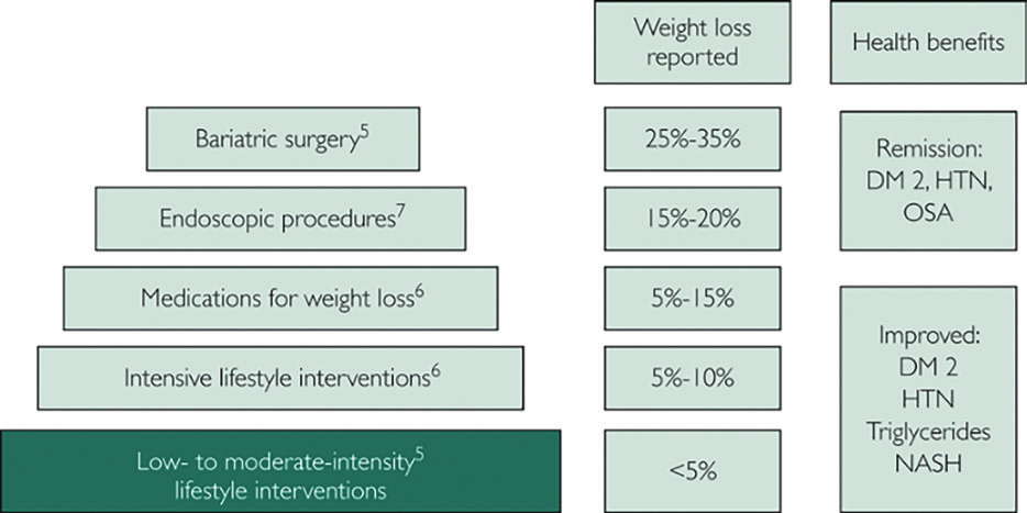
The diet should not only lead to an energy deficit, but also include foods and beverages with antioxidant and anti-inflammatory effects. Stone fruits such as blackberries, raspberries, and blueberries contain flavonoids that reduce inflammation and free oxygen radicals by reducing NF-kB and toll-like receptor 4 (TLR4). Sour cherries are rich in polyphenols and vitamin C and studies have shown that they reduce markers of inflammation and oxidative stress (CRP, IL-6, TNF-α). Another component with antioxidant properties is resveratrol – highly concentrated in grape skins and red wine, respectively. Experimental studies reveal its anti-carcinogenic and anti-inflammatory effects, the ability to reduce weight, hyperglycemia and hyperinsulinemia, protect the heart and endothelial function and prolong life. Omega 3 polyunsaturated fatty acids and high doses of probiotics also show an association with a reduction in BMI and CV risk. (4) Lifestyle changes reduce BMI by 5-10%. Drug treatment contributes to a further 5-15% reduction in BMI. Orlistat, a lipase inhibitor, leads to fat malabsorption and consequently reduces energy intake. Phentermine/topiramate suppresses appetite and reduces food intake through norepinephrine and GABA receptors in the brain.
Naltrexone/bupropion inhibits the reuptake of norepinephrine and dopamine and again suppresses appetite by a central mechanism. Liraglutide 3 mg, a GLP1 receptor agonist, suppresses appetite and delays gastric emptying – a central and peripheral mechanism leading to reduced food intake and showed a significant reduction in MACE and cardiovascular death by 22% in the LEADER study. GLP1 RA reduces appetite and weight, has anti-inflammatory and antiatherosclerotic effects, increases insulin secretion and sensitivity, diuresis and natriuresis, improves quality of life. The next step in the treatment of obesity is endoscopic interventions and bariatric surgery, the results of which are well summarized in the Mayo Clinic success pyramid. (10)
Conclusion
Obesity is a systemic proinflammatory state and through endothelial dysfunction, insulin resistance and neuroendocrine stimulation increases cardiovascular disease and mortality. Obese patients more often suffer from metabolic syndrome, diabetes mellitus, hypertension, coronary artery disease, atrial fibrillation, sleep apnea and heart failure. The risk of SCD is significantly increased. Therefore, obesity should not be considered only as BMI, but mainly in the context of its complications. Modern treatment is successful precisely because it is aimed at reducing cardiovascular risk and includes lifestyle changes as a mandatory and basic element, medications such as GLP1 RA and new endoscopic and surgical procedures.
Bibliography:
- Cardiovascular risk and obesity . C. Cercato1 and F. A. Fonseca (2019) https://doi.org/10.1186/s13098-019-0468-0.
- Metabolically healthy obesity across the life course: epidemiology, determinants, and implications, Catherine M. Phillips, Ann. N.Y. Acad. Sci. 1391 (2017) 85–100 C 2016 New York Academy of Sciences.
- Ectopic Fat Depots and Cardiovascular Disease, Kathryn A. Britton and Caroline S. Fox Circulation. 2011; 124: e837–e841.
- Mechanisms, Pathophysiology and Management of Obesity. Steven B. Heymsfield, M.D., and Thomas A. Wadden, Ph.D. N Engl J Med 2017; 376:254-66.
- Molecular Mechanisms of Obesity-Linked Cardiac Dysfunction: An Up-Date on Current Knowledge. Cells 2021, 10, 629. https://doi.org/10.3390/cells10030629.
- Impact of Obesity-Induced Inflammation on Cardiovascular Diseases (CVD) Gopi Battineni, Getu Gamo Sagaro, etc Int. J. Mol. Sci. 2021, 22, 4798, https://doi.org/10.3390/ijms22094798.
- Obesity and Cardiovascular Disease. A Scientific Statement From the American Heart Association. Circulation. 2021;143:e984–e1010. DOI: 10.1161/CIR.0000000000000973
- Managing Obesity: Scaling the Pyramid to Success Maria L. Collazo-Clavell, MD DOI:https://doi.org/10.1016/j.mayocp.2019.04.011.
- Obesity paradox in cardiovascular disease: where do we stand? Carbone S, Canada JM, Billingsley HE, Siddiqui MS, 2019, https://doi.org/10.2147/VHRM. S168946.
- Diabetes and cardiovascular disease: inter-relation of risk factors and treatment, Aman Sharma. etc, Future Journal of Pharmaceutical Sciences volume 6, Article number: 130 (2020).
- Accuracy of non-invasive blood pressure measurements in obese patients Abigail Arnold, Amanda Mc Naughton Published Online: 11 Jan 2018, https://doi.org/10.12968/bjon.2018.27.1.35.
- Prevalence and characteristics of coronary microvascular dysfunction among chest pain patients in the emergency department. Basmah Safdar 1, Gail D’Onofrio Eur Heart J Acute Cardiovasc Care 2020 Feb; 9 (1): 5-13. doi:10.1177/2048872618764418.Open save panel