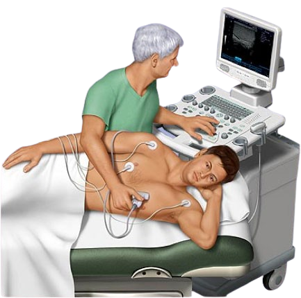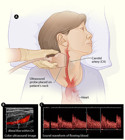
Echocardiography uses ultrasound to examine the structure and function of the heart and great vessels. Ultrasound is used to study the anatomical features of the heart, but also to analyse blood flow and hemodynamics thanks to the Doppler principle. The examination is largely subjective and depends on the capabilities of the ultrasound machine. CW Doppler is used to evaluate valve stenoses. PW Doppler detects pathological blood flow, valve insufficiencies and shunts. Colour Doppler colour-codes blood flow according to direction and average velocity. Tissue Doppler plays a major role in evaluating left ventricular function, myocardial ischemia, distinguishing between constrictive pericarditis and restrictive cardiomyopathy, and evaluating ventricular dyssynchrony. The new Tissue-tracking methods for the assessment of kinetics and function, embedded in the software program of Vivid T8, with which the office is equipped, provide additional information and help in the diagnosis of complicated cases.